All topics
Practice this topic

Strand 1: Organisation of Life

The Cell

Cell organelles, microscopy, and the structure and function of animal and plant cells.

1.3 Unit of Life – Cells

In brief:The cell is the basic unit of life. All living things are made of one or more cells. Animal and plant cells share many organelles but have key differences.

The cell is the basic structural and functional unit of all living things. All cells arise from pre-existing cells (cell theory).

Cell Theory (Schleiden, Schwann, Virchow):
1. All living things are made of one or more cells
2. The cell is the basic unit of life
3. All cells arise from pre-existing cells

Microscopy:
Light microscope: magnification up to ~1,500×. Can view living cells. Uses visible light and glass lenses.
Electron microscope: magnification up to ~500,000×. Much higher resolution. Uses electron beams. Cannot view living cells (specimen must be dead and in a vacuum).

Key differences between plant and animal cells:
Plant cells only: cell wall (cellulose), chloroplasts (photosynthesis), large central vacuole (turgor pressure)
Animal cells only: centrioles (cell division), lysosomes (more prominent), small vacuoles
Both have: cell membrane, cytoplasm, nucleus, mitochondria, ribosomes, ER, Golgi apparatus
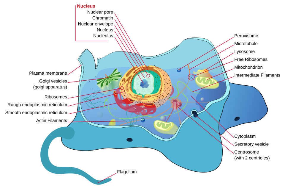
Animal cell

Animal cell

Wikimedia Commons (public domain / CC)
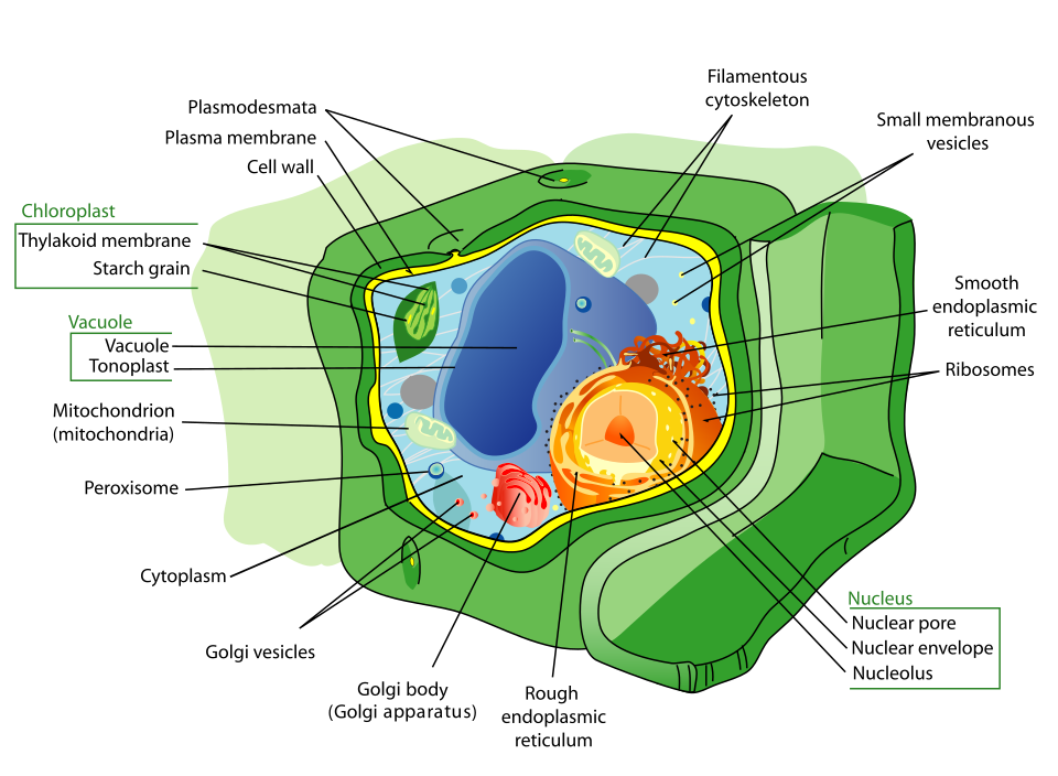
Plant cell

Plant cell

Wikimedia Commons (public domain / CC)

diagram

Key Cell Organelles and Their Functions

NNucleusDNA storage, controls cell
MMitochondrionAerobic respiration (ATP)
CChloroplastPhotosynthesis (plants)
RRibosomeProtein synthesis
GGolgi ApparatusPackages & secretes proteins
EER (Smooth/Rough)Transport, lipid/protein making
CCell WallSupport & protection (plants)
VVacuoleStorage, turgor pressure
comparison

Plant Cell vs Animal Cell

Plant Cell
  • Cell wall (cellulose)
  • Chloroplasts
  • Large central vacuole
  • Regular, fixed shape
  • Starch for energy storage
Animal Cell
  • No cell wall
  • No chloroplasts
  • Small vacuoles
  • Irregular shape
  • Glycogen for energy storage
  • Centrioles present

Key Points

  • 1Levels of organisation: cell → tissue → organ → organ system → organism.
  • 2Light microscope: lower magnification, can view living cells.
  • 3Electron microscope: much higher magnification and resolution, views fine detail of organelles.
  • 4Plant cell only: cell wall, chloroplast, large central vacuole.
  • 5Animal cell only: centrioles (in some), small vacuoles.
  • 6Both: cell membrane, cytoplasm, nucleus, mitochondria, ribosomes, endoplasmic reticulum, Golgi apparatus.

Learning Outcomes

  • Describe the complexity of multicellular organisms
  • Compare the structure of prokaryotic and eukaryotic cells
  • Investigate, using primary data gathered with a light microscope and secondary data gathered from scanning electron microscope imagery, the structures and organelles of animal and plant cells and relate them to their functions

2. Using the Microscope: Magnification, Resolution and Cell Size

In brief:The microscope is the tool that made cell biology possible; you need to be able to prepare a slide and calculate magnification.

Magnification is how many times larger the image is than the object. Resolution is the smallest distance between two points that can still be seen as separate. Increasing magnification without resolution just gives a bigger blur, which is why the electron microscope, using a much shorter electron wavelength, reveals detail the light microscope never can.

Calculations

Total magnification = eyepiece × objective. A ×10 eyepiece with a ×40 objective gives ×400.

Actual size = image size ÷ magnification. Keep units consistent: 1 mm = 1000 µm.

Worked example: a cell measures 20 mm on a photograph taken at ×400. Actual size = 20 ÷ 400 = 0.05 mm = 50 µm.

Preparing a slide of onion or cheek cells

  1. Place a drop of water on a clean slide.
  2. Peel a single layer of epidermis from an onion (or gently scrape the inside of the cheek) and lay it flat in the drop.
  3. Add a drop of stain: iodine for plant cells, methylene blue for animal cells. Stain increases contrast so the nucleus becomes visible.
  4. Lower the coverslip at an angle with a mounting needle. This avoids trapping air bubbles, which look like thick black circles.
  5. Focus on low power first using the coarse focus, then switch to high power and use the fine focus only.

Typical sizes

Plant cell 50–100 µm, animal cell 10–30 µm, bacterium 1–5 µm, virus 20–300 nm, ribosome about 25 nm. Only the first three are visible with a school light microscope.

svg

Light microscope vs electron microscope

Light (compound) microscope Uses light and glass lenses Magnification up to about ×1500 Living or stained material, in colour Shows cell wall, nucleus, chloroplasts Cheap, portable, used in school labs Electron microscope Uses a beam of electrons and magnets Magnification over ×500 000 Dead material only, in a vacuum Reveals ultrastructure: ribosomes, cristae Large, expensive, black and white images Total magnification = eyepiece lens × objective lens ×10 eyepiece with a ×40 objective gives ×400. Image size = actual size × magnification. 1 mm = 1000 µm. A typical plant cell is 50–100 µm; a bacterium is 1–5 µm.

Key Points

  • 1Total magnification = eyepiece lens x objective lens
  • 2Actual size = image size / magnification; 1 mm = 1000 micrometres
  • 3Resolution, not magnification, limits the detail a microscope can show
  • 4Iodine stains plant cells, methylene blue stains animal cells
  • 5Lower the coverslip at an angle to avoid air bubbles; focus on low power first

Learning Outcomes

  • Prepare and examine an animal cell and a plant cell using a light microscope
  • Calculate magnification and actual cell size from a microscope image

3. Comparing Animal, Plant, Bacterial and Fungal Cells

In brief:A single comparison you can use for any cell-identification question in the exam.

All cells share a cell membrane, cytoplasm, ribosomes and DNA. Everything else varies.

FeatureAnimalPlantBacterialFungal
NucleusYesYesNo - free loop of DNA in the cytoplasmYes, often several per cell
Cell wallNoneCellulosePeptidoglycanChitin
ChloroplastsNoYesNoNo
VacuoleSmall, temporaryOne large permanent vacuoleNoneSmall
MitochondriaYesYesNoYes
Ribosomes80S (larger)80S70S (smaller)80S
NutritionHeterotrophicAutotrophicBoth types occurHeterotrophic, saprophytic or parasitic

Prokaryotic or eukaryotic?

Prokaryotic cells (bacteria and archaea) have no nucleus and no membrane-bound organelles. Their DNA is a single circular chromosome, often with small extra rings called plasmids. They are small, they have 70S ribosomes and they divide by binary fission.

Eukaryotic cells (animals, plants, fungi, protists) have a true nucleus enclosed by a nuclear membrane, membrane-bound organelles, linear chromosomes wound around proteins, 80S ribosomes, and they divide by mitosis or meiosis.

Why bacterial ribosomes matter

Antibiotics such as streptomycin bind to the 70S bacterial ribosome and block protein synthesis, but leave our 80S ribosomes alone. That difference is what makes an antibiotic safe to take.

svg

Four cell types side by side

Four cell types compared. Only the bacterial cell is prokaryotic. Animal cell no wall, no chloroplast small vacuoles, centrioles Plant cell cellulose wall, chloroplasts one large vacuole Bacterial cell no nucleus, loop of DNA peptidoglycan wall, flagellum Fungal hypha chitin wall, many nuclei no chloroplasts, saprophytic

Key Points

  • 1All cells have a membrane, cytoplasm, ribosomes and DNA
  • 2Cell walls: cellulose in plants, peptidoglycan in bacteria, chitin in fungi, none in animals
  • 3Prokaryotes have no nucleus and no membrane-bound organelles; DNA is a circular loop plus plasmids
  • 4Bacterial ribosomes are 70S, eukaryotic are 80S, which is why some antibiotics are selective
  • 5Only plant cells have chloroplasts and a single large permanent vacuole

Learning Outcomes

  • Compare the structure of animal, plant, bacterial and fungal cells
  • Distinguish between prokaryotic and eukaryotic cells