All topics
Practice this topic

Strand 2: Structures & Processes

Response Systems

The musculoskeletal system, nervous system, neurotransmission, the endocrine system, and the immune system.

2.4 The Musculoskeletal System

In brief:The skeleton provides support, protection, and movement. Muscles work in antagonistic pairs at joints.

The Human Skeleton

The skeleton is divided into two parts:

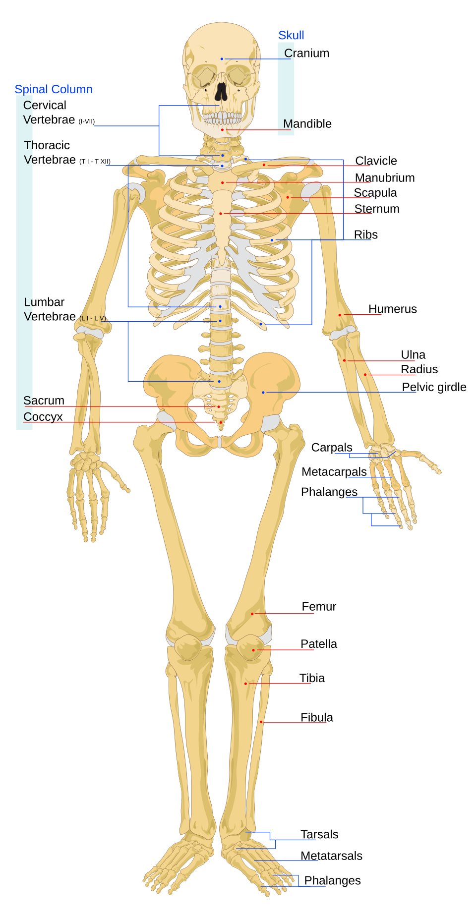
Axial Skeleton (central axis):

  • Skull: protects the brain
  • Vertebrae: protect the spinal cord; intervertebral discs between vertebrae act as shock absorbers and allow flexibility
  • Ribs (12 pairs): protect the heart and lungs
  • Sternum (breastbone): protects the heart, attachment point for ribs

Appendicular Skeleton (limbs and girdles):

  • Pectoral girdle: clavicle (collarbone) + scapula (shoulder blade) - attaches arms to axial skeleton
  • Pelvic girdle: supports lower body weight, protects reproductive organs
  • Upper limb: humerus (upper arm) → radius + ulna (forearm)
  • Lower limb: femur (thigh) → patella (kneecap) → tibia + fibula (shin)

Synovial Joints

Moveable joints with the following structures:

  • Cartilage: smooth, slippery tissue on bone ends - reduces friction, absorbs shock
  • Ligaments: tough, elastic connective tissue - bone to bone, holds joint together
  • Tendons: strong, inelastic connective tissue - muscle to bone, transmits force
  • Synovial fluid: lubricates the joint, reduces friction
  • Synovial membrane: produces synovial fluid

Antagonistic Muscle Pairs

Muscles can only pull (contract), not push. Therefore they work in antagonistic pairs:

  • Biceps contracts → arm bends (flexion). Triceps relaxes.
  • Triceps contracts → arm straightens (extension). Biceps relaxes.
Human skeleton

Human skeleton

Wikimedia Commons (public domain / CC)

Immune response

Overview of the innate and adaptive immune response

Wikimedia Commons (CC)

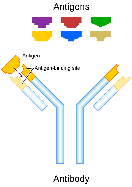
Antibody structure

Structure of an antibody (immunoglobulin G)

Wikimedia Commons (CC)

Key Points

  • 1Axial skeleton: skull, vertebrae, ribs, sternum. Protects brain, spinal cord, heart, lungs.
  • 2Appendicular skeleton: pectoral girdle (clavicle + scapula), pelvic girdle, upper and lower limbs.
  • 3Intervertebral discs act as shock absorbers between vertebrae.
  • 4Synovial joints: cartilage (reduces friction), ligaments (bone-bone), tendons (muscle-bone), synovial fluid (lubricates).
  • 5Antagonistic muscle pairs: biceps/triceps - one contracts while the other relaxes.

Learning Outcomes

  • Outline the structures and systems for response in humans and plants
  • Relate the structures of the component parts of the axial and appendicular skeleton to their functions
  • Model the function of an antagonistic muscle pair; relate the functions of cartilage, ligament and tendons in synovial joints

The Nervous System

In brief:The nervous system coordinates rapid responses to stimuli. It consists of the CNS (brain and spinal cord) and PNS (peripheral nerves), using electrical impulses and chemical neurotransmitters.

Structure of the Nervous System

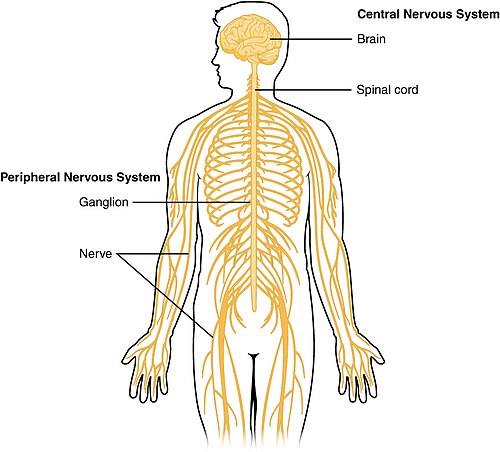
Central Nervous System (CNS): brain + spinal cord - processes information and coordinates responses.
Peripheral Nervous System (PNS): all nerves outside the CNS - sensory nerves carry impulses to the CNS, motor nerves carry impulses from the CNS.

A nerve is a bundle of nerve cells (neurons) grouped together, similar to individual wires in electrical cabling. Nerve cells do not undergo mitosis - if damaged, they cannot be replaced.

Neuron Structure

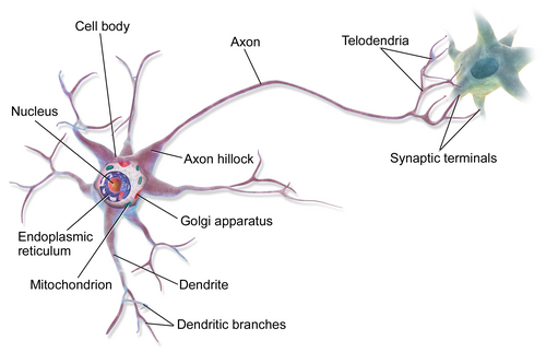
Each neuron has three main parts:

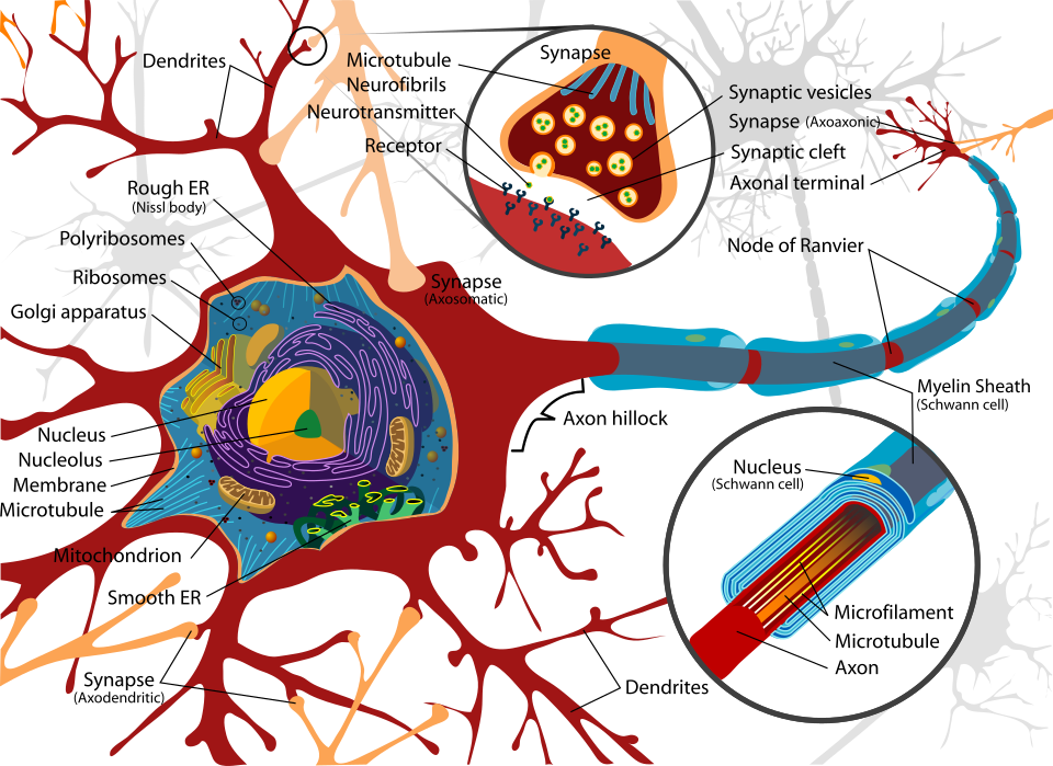
  • Dendrites: short branching fibres that convert chemical messages to electrical impulses and direct them toward the cell body. Branched to increase surface area for receiving impulses.
  • Cell body (soma): contains the nucleus and organelles. Produces neurotransmitter chemicals.
  • Axon: long fibre that conducts electrical impulses away from the cell body.
  • Schwann cells: wrap around the axon to form the myelin sheath.
  • Myelin sheath: white fatty insulating layer that protects and electrically insulates the axon, speeding up conduction of impulses.
  • Nodes of Ranvier: gaps in the myelin sheath where ions move in/out, enabling saltatory conduction (impulse 'jumps' between nodes).
  • Synaptic knob: terminal endpoint of the axon containing neurotransmitter vesicles that store chemical messengers.

Types of Neurons

  • Sensory neurons: carry impulses from receptors to the CNS. Cell body is on a side-branch off the axon.
  • Motor neurons: carry impulses from the CNS to effectors (muscles/glands). Cell body at the end of the axon.
  • Interneurons (relay neurons): connect sensory and motor neurons within the CNS. Cell body located midway along the axon.

The Brain - Structure and Function

The brain is protected by the skull. It is surrounded by three membranes called the meninges and cerebrospinal fluid (a clear liquid that protects against shock and provides nutrients).

The CNS has two regions: white matter (axons - carries impulses) and grey matter (cell bodies - processes information). In the brain, grey matter is on the outside; in the spinal cord, grey matter forms a butterfly shape in the centre.

Brain StructureFunction
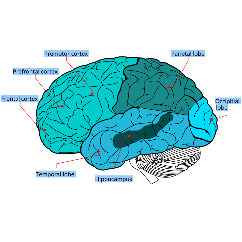
Cerebral cortex (cerebrum)Receives and processes impulses from sense organs. Controls memory, language, voluntary muscle movement, and intelligence.
CerebellumControls unconscious motor functions: muscle coordination and balance.
Brain stemControls involuntary actions: heart rate, breathing rate, blood pressure.
ThalamusRelay station - all motor and sensory information passes through before being directed to the cerebral cortex for interpretation.
EpithalamusContains the pineal gland which secretes melatonin to control circadian (24-hour) rhythms. Regulates motor pathways and emotions.
HypothalamusControls homeostasis - body temperature, fluid balance, metabolism. Links nervous and endocrine systems by controlling the pituitary gland.

The Spinal Cord

Controls most reflex actions. 31 pairs of spinal nerves connect the spinal cord to specific body regions. Each spinal nerve branches into:

  • Dorsal root (back): carries sensory neurons to the spinal cord. Cell bodies lie in the dorsal root ganglion.
  • Ventral root (front): carries motor neurons from the spinal cord.

A central canal filled with cerebrospinal fluid runs through the centre.

Neurotransmission - Mechanism of Impulse Transfer

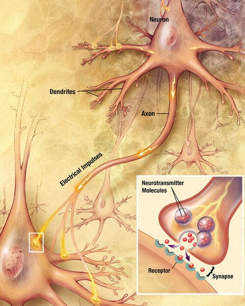
Nerve impulses are electro-chemical: electrical along the axon, chemical across synapses.

  1. At rest, the neuron membrane has a charge difference: inside negative, outside positive.
  2. A threshold stimulus (minimum strength required) must be reached for an impulse to fire. Below threshold → no impulse.
  3. All-or-nothing rule: once threshold is reached, the same impulse is carried regardless of stimulus strength.
  4. Movement of ions reverses the charge (inside becomes positive) - this change travels along the axon to the synaptic knob.
  5. At the synapse: impulse triggers neurotransmitter vesicles to release chemicals (e.g. acetylcholine) into the synaptic cleft.
  6. Neurotransmitters diffuse across the cleft and lock into receptors on the post-synaptic neuron.
  7. This causes an inrush of ions → new impulse generated in the next neuron.
  8. Enzymes in the synaptic cleft digest the neurotransmitters, stopping the response. Products are recycled by the pre-synaptic neuron.
  9. The neuron returns to its resting charges. The refractory period is the minimum time required before the neuron can carry another impulse.

The Reflex Arc

A reflex action is a rapid, involuntary, automatic response. The impulse bypasses the brain (goes only to the spinal cord) for faster protection:

  1. Sensory receptor detects a stimulus (e.g. pain receptor in skin)
  2. Sensory neuron carries impulse to spinal cord via dorsal root
  3. Sensory neuron synapses with an interneuron in the CNS
  4. Interneuron synapses with a motor neuron
  5. Motor neuron carries impulse out via ventral root to an effector muscle
  6. Muscle contracts → response (e.g. hand pulled away from hot object)

The brain can sometimes override a reflex (e.g. holding a hot plate to avoid dropping food) - but this must be learned.

Enzyme Inhibition at Synapses

Some medicines inhibit enzymes that break down neurotransmitters, increasing their levels and prolonging the response:

  • Alzheimer's treatment: enzyme inhibitors enhance acetylcholine levels, improving nerve cell communication.
  • SSRIs (for depression): block serotonin reuptake, keeping serotonin active longer.
  • Nicotine: inhibits enzymes that break down neurotransmitters → overstimulation of receptors. Also raises dopamine → addiction.

Dopamine & Endorphins

Endorphins: neurotransmitters released by the pituitary gland and hypothalamus during pleasurable activities (eating, laughing, exercise). Known as the body's natural painkillers - they block nerves receiving pain signals, allowing the body to function in stressful situations.

Dopamine: the 'feel-good' neurotransmitter released following pleasurable activities. Creates a temporary 'dopamine rush' - explains why sugary foods are addictive. When the brain doesn't produce enough dopamine → Parkinson's disease (involuntary shaking), treated with L-dopa (converted to dopamine in the body).

Dopamine and endorphins work together: e.g. a runner experiences muscle pain → endorphins released to block pain → followed by a dopamine surge → 'runner's high' → encourages running again.

Addictive drugs (e.g. cocaine) interfere with normal dopamine recycling, allowing excessive dopamine to remain active.

Lifestyle factors: exercise (↑ both), dietary choices, sleep quality, social interaction all affect dopamine and endorphin levels.

Homeostasis

Homeostasis = maintaining a stable internal environment (e.g. body temperature, blood pH, water content, CO₂ levels). Uses negative feedback: change detected → response opposes change → returns to normal.

Components: receptors (detect change) → control centre (CNS/hypothalamus) → effectors (muscles/glands carry out response).
Neuron - dendrites, axon, myelin

Neuron - dendrites, axon, myelin

Wikimedia Commons (public domain / CC)

Lobes of the brain

Lobes of the brain

Wikimedia Commons (public domain / CC)

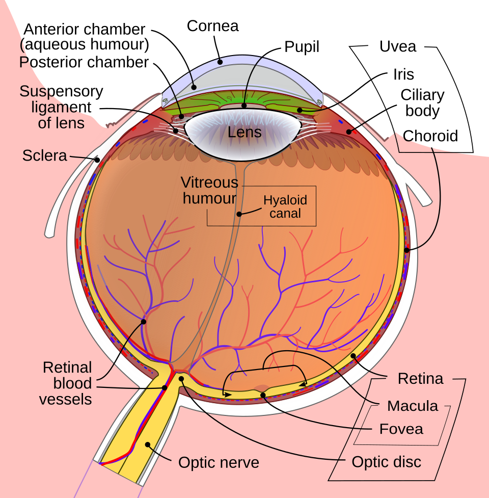
The human eye

The human eye

Wikimedia Commons (public domain / CC)

table

Parts of the Brain

StructureFunction
Cerebral cortexMemory, language, voluntary movement, intelligence
CerebellumMuscle coordination and balance
Brain stemHeart rate, breathing, blood pressure (involuntary)
ThalamusRelay station - directs sensory/motor info to cortex
EpithalamusPineal gland → melatonin → circadian rhythms
HypothalamusHomeostasis - temperature, fluid balance, links to pituitary
process

Neurotransmission Across a Synapse

Threshold stimulus reached
Ion movement reverses charge along axon
Vesicles release neurotransmitter into cleft
Neurotransmitter binds to post-synaptic receptors
New impulse in next neuron
Enzymes digest & recycle neurotransmitter
process

The Reflex Arc

Stimulus
Receptor
Sensory Neuron
(dorsal root)
Interneuron
(spinal cord)
Motor Neuron
(ventral root)
Effector
comparison

Nervous vs Hormonal Control

Nervous System
  • Fast response (milliseconds)
  • Electrical impulses via neurons
  • Short-lasting effect
  • Targets specific muscles/glands
  • Response is precise
Hormonal System
  • Slow response (seconds to hours)
  • Chemical messengers via blood
  • Long-lasting effect
  • Targets widespread organs
  • Response is more general

Key Points

  • 1CNS = brain + spinal cord. PNS = sensory + motor nerves. Nerve cells cannot undergo mitosis.
  • 2Neuron parts: dendrites, cell body (soma), axon, myelin sheath (Schwann cells), nodes of Ranvier, synaptic knob.
  • 3Brain: cerebral cortex (voluntary), cerebellum (coordination), brain stem (involuntary), thalamus (relay), hypothalamus (homeostasis), epithalamus (circadian rhythms).
  • 4Spinal cord: 31 pairs of spinal nerves. Dorsal root = sensory in. Ventral root = motor out. Grey matter butterfly-shaped in centre.
  • 5Threshold stimulus required for impulse. All-or-nothing rule: same impulse regardless of stimulus strength.
  • 6Synapse: neurotransmitters released → bind receptors → enzymes digest → products recycled. Refractory period before next impulse.
  • 7Reflex arc bypasses brain → faster response via spinal cord. Brain can override learned reflexes.
  • 8Enzyme inhibitors increase neurotransmitter levels (Alzheimer's treatment, SSRIs, nicotine mechanism).
  • 9Endorphins: natural painkillers from pituitary/hypothalamus. Dopamine: reward neurotransmitter. Low dopamine → Parkinson's (treated with L-dopa).
  • 10Homeostasis: negative feedback - receptors → control centre → effectors → return to normal.

Learning Outcomes

  • Relate the structure of the parts of the central nervous system and the peripheral nervous system to their functions
  • Compare nervous and hormonal coordination
  • Relate the structures of a motor and sensory neuron to their functions
  • Explain the role of neurotransmitters at a synapse
  • Model impulse travel across a synaptic cleft, consider the impacts of disruptions to impulse travel
  • Discuss the roles of the neurotransmitters dopamine and endorphins in humans, taking into account the influence of lifestyle choices on their levels in the human body

The Endocrine System

In brief:The endocrine system uses hormones (chemical messengers) secreted by glands to coordinate slower, longer-lasting body responses.

The Endocrine System

The endocrine system consists of glands that secrete hormones (chemical messengers) directly into the blood. Hormones travel via the bloodstream to target organs where they produce a response.

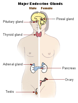
Major Endocrine Glands

GlandLocationHormone(s)Function
PituitaryBase of brainFSH, LH, oxytocin, TSH, ADH'Master gland' - controls other glands
ThyroidNeckThyroxineControls metabolic rate
ParathyroidBehind thyroidParathyroid hormoneRaises blood calcium levels
AdrenalAbove kidneysAdrenalineFight or flight response
PancreasBehind stomachInsulinLowers blood glucose
TestesScrotumTestosteroneMale secondary sexual characteristics, sperm production
OvariesPelvic areaOestrogen, ProgesteroneFemale secondary sexual characteristics, menstrual cycle

Hormonal Manipulations

  • Sport: anabolic steroids (synthetic testosterone) used illegally to build muscle - causes liver damage, heart problems
  • Health: insulin injections for diabetes, HRT (hormone replacement therapy) for menopause, contraceptive pill
  • Agriculture: growth hormones in livestock, plant growth regulators
Endocrine glands

Endocrine glands

Wikimedia Commons (public domain / CC)

Key Points

  • 1Pituitary gland: 'master gland' - secretes FSH, LH, oxytocin, TSH, ADH.
  • 2Thyroid: thyroxine (metabolic rate). Parathyroid: parathyroid hormone (blood calcium).
  • 3Adrenal glands: adrenaline (fight or flight). Pancreas: insulin (lowers blood glucose).
  • 4Ovaries: oestrogen + progesterone. Testes: testosterone.
  • 5Hormonal manipulations: sport (anabolic steroids), health (insulin, HRT), agriculture (growth hormones).

Learning Outcomes

  • Compare nervous and hormonal coordination
  • Identify the location of the major glands in the endocrine system, describe the functions of their associated hormones
  • Recognise the impact of hormonal manipulations on organisms

The Immune System

In brief:The immune system defends the body against pathogens using innate (non-specific) and adaptive (specific) responses.

Pathogens

Disease-causing organisms: prions, bacteria, protists, viruses, fungi, parasitic animals.

Innate Immunity (Non-Specific)

Immediate, does not improve with repeated exposure:

  • Physical barriers: skin, mucus membranes, stomach acid, tears (lysozyme)
  • Phagocytes (e.g. monocytes): engulf and destroy pathogens by phagocytosis
  • Natural killer cells: destroy virus-infected cells and cancer cells
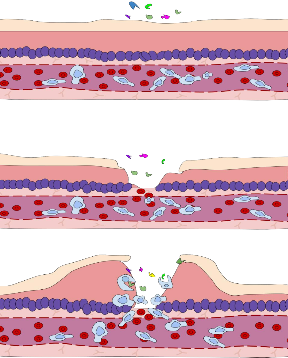
  • Inflammation: increased blood flow brings white blood cells to infection site

Adaptive Immunity (Specific)

Slower to develop but has memory for faster future responses:

  • B lymphocytes: produce antibodies (Y-shaped proteins) that bind to specific antigens on the pathogen surface → neutralise them
  • T lymphocytes:
    • Helper T cells: activate B cells and other immune cells
    • Killer T cells: destroy infected cells directly
    • Suppressor T cells: switch off immune response when infection cleared
    • Memory T cells: provide long-term immunity

Acquired Immunity

  • Active immunity: body makes its own antibodies - by infection or vaccination (weakened/inactive pathogen triggers memory cell production)
  • Passive immunity: antibodies received from another source - breast milk (colostrum), antibody injections

Viral Replication

Viruses cannot reproduce on their own - they hijack host cells:

  1. Virus attaches to host cell surface
  2. Injects genetic material (DNA or RNA) into cell
  3. Host cell machinery copies viral genes and makes viral proteins
  4. New virus particles assembled
  5. Host cell bursts (lysis), releasing new viruses

Infectious Disease Spread

Emergence and spread depends on: persistence in host, mutations, antibiotic resistance, population immunity, mobility of populations, R₀ (reproduction number - average number of people one infected person infects).

Key Points

  • 1Pathogens: prions, bacteria, protists, viruses, fungi, parasitic animals.
  • 2Innate immunity: non-specific, immediate - skin, mucus, phagocytes, inflammation, natural killer cells.
  • 3Adaptive immunity: specific, has memory - B lymphocytes (antibodies) and T lymphocytes.
  • 4T cell types: helper (activates), killer (destroys), suppressor (stops), memory (long-term).
  • 5Active immunity: own antibodies (infection or vaccination). Passive immunity: received antibodies.
  • 6Viruses replicate inside host cells - inject genetic material, hijack cell machinery, new viruses released.
  • 7R₀ value: average number infected by one case. Factors: mutations, resistance, population immunity.

Learning Outcomes

  • Distinguish between innate and acquired immunity; outline the strategies applied to prevent and treat microbial diseases
  • Model how viruses replicate within cells
  • Compare the roles of different types of white blood cell in immune response
  • Explore factors that contribute to the emergence of infectious diseases in plants and animals
  • Discuss the importance of a knowledge of emerging diseases in society

4. The Nervous System

In brief:The nervous system gives fast, short-lived responses to changes in the environment through electrical impulses in neurons.

A stimulus is any change in the environment. A receptor detects it, the CNS processes the information, and an effector (muscle or gland) carries out the response. The nervous system works with the hormonal system to maintain homeostasis.

Two divisions

  • Central Nervous System (CNS) - brain and spinal cord
  • Peripheral Nervous System (PNS) - all the nerves that carry impulses to and from the CNS

Neurons (nerve cells)

Made of dendrites (receive impulses), a cell body with the nucleus, and a long axon (carries the impulse). The axon is often insulated by a myelin sheath broken by nodes of Ranvier, which lets impulses jump and travel faster. Mature neurons do not divide by mitosis, so damage is often permanent.

TypeJobCell body position
Sensory neuronReceptor → CNSSide branch off axon
Inter-neuronWithin the CNSMiddle of axon
Motor neuronCNS → effectorEnd of axon

Impulse & synapse

At rest the inside of an axon is negatively charged. A stimulus at or above the threshold lets Na⁺ ions rush in, flipping the charge and creating a nerve impulse that travels along the axon. The refractory period is the short time before the neuron can fire again.

Where two neurons meet is a synapse. The impulse cannot jump the gap electrically, so neurotransmitter chemicals stored in vesicles are released into the synaptic cleft. They bind to receptors on the next neuron and start a new impulse. Enzymes then break the neurotransmitter down so the signal stops. Examples of neurotransmitters: dopamine (reward, movement) and endorphins (block pain signals - the body's natural painkiller).

Reflex arc

A very fast, automatic response that does not need the brain (e.g. pulling your hand off a hot plate). Pathway: receptor → sensory neuron → inter-neuron in spinal cord → motor neuron → effector. The brain is informed a fraction later.

The brain

PartMain role
Cerebral cortexThought, memory, voluntary movement, senses
CerebellumBalance and coordinated movement
ThalamusRelay station for sensory information
HypothalamusHomeostasis - temperature, water, hunger
Brain stem (incl. medulla)Automatic controls - heart rate, breathing

The CNS is protected by bone (skull, vertebrae), three meninges membranes and cerebrospinal fluid that cushions and feeds it. Grey matter = cell bodies; white matter = myelinated axons.

Labelled motor neuron

Structure of a motor neuron - dendrites, cell body, axon, myelin

Blausen Medical / Wikimedia Commons (CC BY)

Chemical synapse

Chemical synapse - neurotransmitter crosses the synaptic cleft

Wikimedia Commons (CC BY-SA)

Central and peripheral nervous system

The CNS (brain and spinal cord) and the PNS (peripheral nerves)

OpenStax / Wikimedia Commons (CC BY)

Key Points

  • 1Stimulus → receptor → CNS → effector → response
  • 2CNS = brain + spinal cord; PNS = all other nerves
  • 3Three neuron types: sensory, inter-neuron, motor
  • 4Impulse travels along axon; neurotransmitter crosses the synapse
  • 5Reflex arc bypasses the brain for speed
  • 6Endorphins block pain; dopamine linked to reward
  • 7Brain parts: cerebral cortex, cerebellum, thalamus, hypothalamus, brain stem