All topics
Practice this topic

Strand 2: Structures & Processes

Transport & Transfer

Membrane transport, the urinary system, digestion, the breathing system, the circulatory system, and transport in plants.

2.6 Membrane Transport

In brief:Materials move across cell membranes by diffusion, osmosis, and active transport.

Materials must cross the cell membrane (phospholipid bilayer) to enter or exit cells. Three main methods:

1. Diffusion (Passive - no energy)

  • Movement of molecules from high → low concentration (down the gradient)
  • Examples: O₂ into cells, CO₂ out of cells, gas exchange in alveoli
  • Factors affecting rate: concentration gradient, temperature, surface area, membrane thickness

2. Osmosis (Special case of diffusion)

  • Movement of water from a dilute solution → concentrated solution through a semi-permeable membrane
  • Plant cell in water: water enters vacuole → cell swells → becomes turgid (rigid - important for plant support)
  • Plant cell in concentrated solution: water leaves → cell shrinks → plasmolysis (membrane pulls away from wall)
  • Animal cell in water: water enters → cell may burst (lysis)
  • Animal cell in concentrated solution: water leaves → cell shrivels (crenation)
  • Food preservation: salt/sugar creates concentrated solution outside cells → water leaves microorganisms by osmosis → they die/cannot grow

3. Active Transport (Requires ATP energy)

  • Movement of molecules against the concentration gradient (low → high)
  • Uses carrier proteins and energy from ATP
  • Examples: mineral uptake by root hair cells, glucose absorption in gut

Key Points

  • 1Diffusion: passive movement from high to low concentration. No energy needed.
  • 2Osmosis: water moves from dilute to concentrated solution through a semi-permeable membrane.
  • 3Active transport: movement against gradient, requires ATP and carrier proteins.
  • 4Turgor: plant cells swell with water and become rigid (important for support).
  • 5Food preservation: salt/sugar draws water out of microorganisms by osmosis.

Learning Outcomes

  • Distinguish between diffusion, osmosis and active transport; examine the role of osmosis in food preservation and plant health
  • Investigate factors affecting rates of osmosis across semi-permeable membranes, use primary data to support conclusions

The Urinary System

In brief:The urinary system filters blood, removes waste, and regulates water balance.

Urinary System Structure

Components: kidneys (×2) → uretersurinary bladderurethra

Kidney Structure

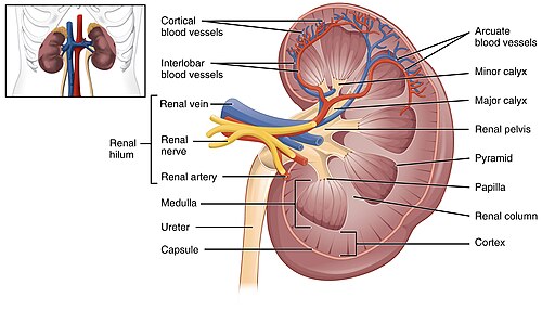
  • Cortex: outer region containing Bowman's capsules and convoluted tubules
  • Medulla: inner region containing loops of Henle and collecting ducts
  • Renal pelvis: funnel-shaped area collecting urine, drains into ureter
  • Renal artery: brings blood to be filtered
  • Renal vein: carries filtered blood away

The Nephron (HL)

The functional unit of the kidney (~1 million per kidney):

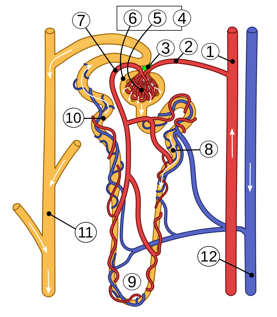
  1. Bowman's capsule + glomerulus: blood enters glomerulus at high pressure → small molecules (water, glucose, amino acids, urea, salts) are filtered into Bowman's capsule. Large molecules (proteins, blood cells) stay in blood.
  2. Proximal convoluted tubule: most reabsorption occurs - glucose, amino acids, and most water/salts are actively transported back into blood
  3. Loop of Henle: creates a concentration gradient in the medulla → enables water reabsorption
  4. Distal convoluted tubule: fine-tuning of salt and water balance
  5. Collecting duct: remaining fluid (urine) passes down to renal pelvis

Osmoregulation - Role of ADH

ADH (Antidiuretic Hormone) from the pituitary gland controls water reabsorption:

  • Dehydrated (blood too concentrated): more ADH released → collecting duct becomes more permeable → more water reabsorbed → small volume of concentrated urine
  • Over-hydrated (blood too dilute): less ADH released → collecting duct less permeable → less water reabsorbed → large volume of dilute urine
Nephron structure (kidney)

Nephron structure (kidney)

Wikimedia Commons (public domain / CC)

Key Points

  • 1Urinary system: kidneys → ureters → bladder → urethra.
  • 2Kidney regions: cortex (outer), medulla (inner), renal pelvis (collects urine).
  • 3Nephron: Bowman's capsule (filtration) → proximal tubule (reabsorption) → Loop of Henle → distal tubule → collecting duct.
  • 4Glomerular filtration: high pressure forces small molecules into Bowman's capsule.
  • 5ADH controls water reabsorption: more ADH = more water reabsorbed = concentrated urine.

Learning Outcomes

  • Relate the macrostructure of the urinary system to its function in filtering and removing waste; outline the filtration of blood in the nephron

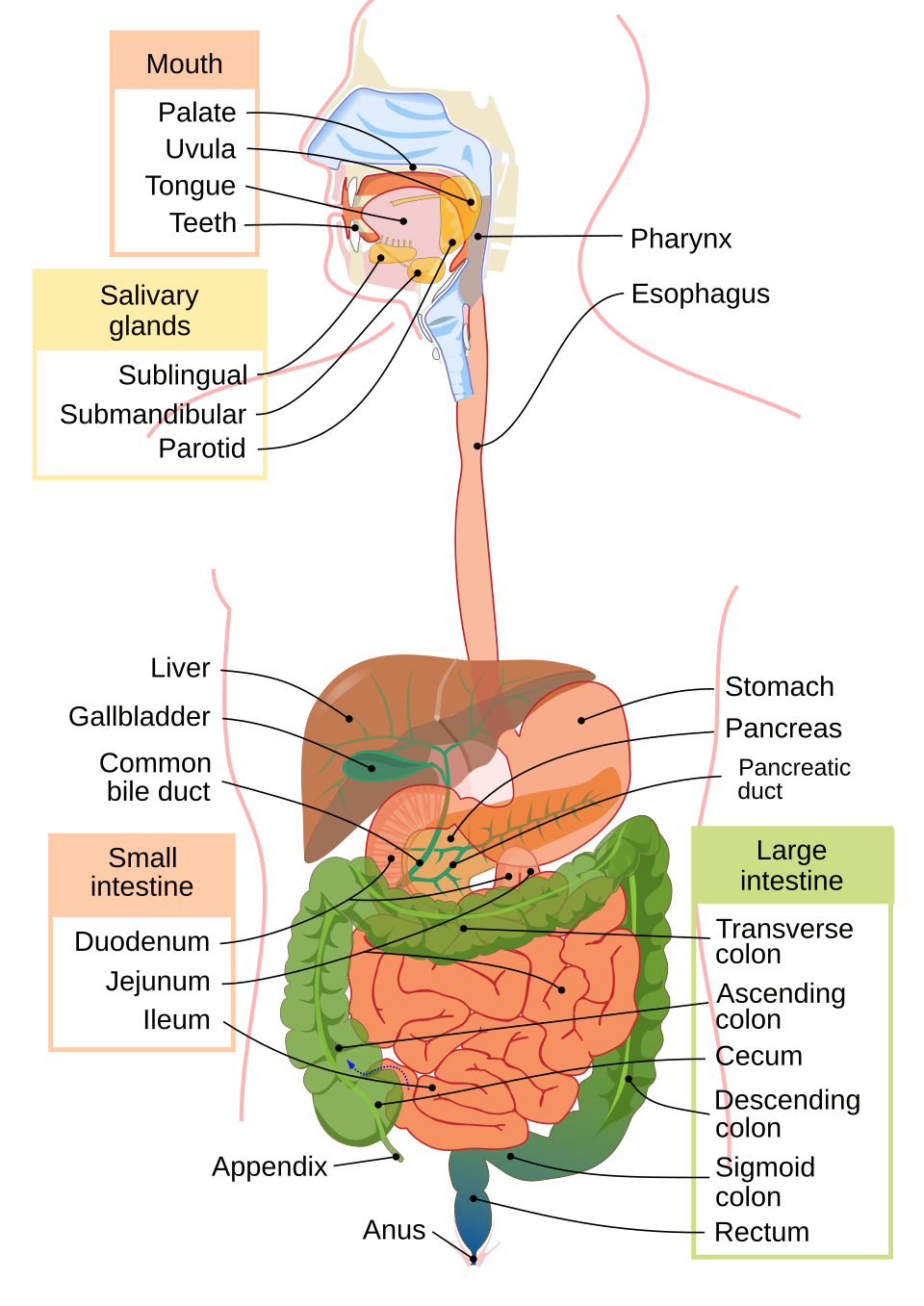
The Digestive System

In brief:The digestive system breaks down food mechanically and chemically so nutrients can be absorbed into the blood.

The digestive system breaks down food into small soluble molecules for absorption.

Four Stages

IngestionDigestionAbsorptionEgestion

Mechanical Digestion

  • Teeth: chewing breaks food into smaller pieces
  • Peristalsis: wave-like muscle contractions push food along the tract
  • Stomach churning: muscular walls mix food with gastric juice
  • Bile: emulsifies fats into smaller droplets (increases surface area for lipase)

Chemical Digestion

LocationEnzymeSubstrate → ProductpH
MouthSalivary amylaseStarch → Maltose~7 (neutral)
StomachPepsin (protease)Protein → Peptides~2 (HCl)
Small intestinePancreatic amylaseStarch → Maltose~8 (alkaline)
Small intestineTrypsin (protease)Protein → Amino acids~8
Small intestineLipaseFats → Fatty acids + Glycerol~8

Absorption

Nutrients absorbed through villi in the small intestine. Villi adaptations: finger-like projections (large surface area), one cell thick (short diffusion distance), rich blood supply (maintains gradient), lacteals (absorb fats).

Hepatic portal vein carries absorbed nutrients to the liver for processing: glycogen storage, deamination of excess amino acids (produces urea).

Large Intestine

Absorbs water from undigested material. Remaining waste stored in rectum and eliminated via anus (egestion).

Balanced Diet

A balanced diet includes: carbohydrates, proteins, fats, vitamins, minerals, water, and dietary fibre. Requirements vary with age, gender, and activity level.

Human digestive system

Human digestive system

Wikimedia Commons (public domain / CC)

table

Digestive Enzymes Summary

EnzymeTypeSubstrate → ProductWhere ProducedWhere Active
Salivary amylaseCarbohydraseStarch → MaltoseSalivary glandsMouth (pH 7)
PepsinProteaseProtein → PeptidesStomach wallStomach (pH 2)
Pancreatic amylaseCarbohydraseStarch → MaltosePancreasSmall intestine
TrypsinProteaseProtein → Amino acidsPancreasSmall intestine
LipaseLipaseFats → Fatty acids + GlycerolPancreasSmall intestine
Bile (not enzyme)-Emulsifies fatLiver (stored in gallbladder)Small intestine

Key Points

  • 1Four stages: ingestion, digestion (mechanical + chemical), absorption, egestion.
  • 2Mechanical: teeth, peristalsis, stomach churning, bile emulsification.
  • 3Chemical: amylase (starch→maltose), protease (protein→amino acids), lipase (fats→fatty acids+glycerol).
  • 4Absorption through villi in small intestine - large surface area, rich blood supply, one cell thick.
  • 5Hepatic portal vein → liver: glycogen storage and deamination of amino acids.
  • 6Large intestine absorbs water. Balanced diet varies with age, gender, activity.

Learning Outcomes

  • Model how the macrostructure of the human digestive system and associated organs and glands carry out the process of digesting fats, carbohydrates and proteins
  • Describe the absorption, transport and storage of the products of digestion
  • Research the biological implications of dietary choices

The Breathing System

In brief:The breathing system facilitates gas exchange between the body and the external environment.

Structure of the Breathing System

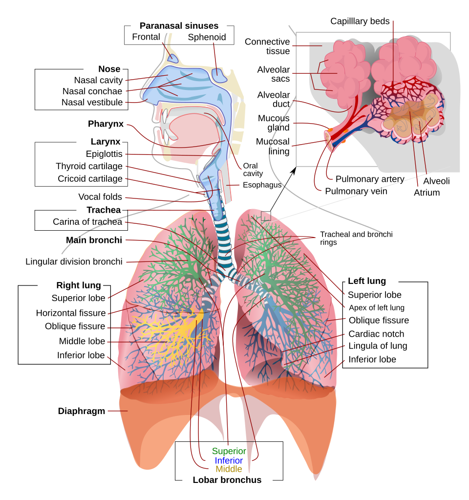
Breathing tract: nose/mouth → pharynx → larynx → trachea (lined with C-shaped cartilage rings to keep it open) → bronchi (one to each lung) → bronchiolesalveoli

Gas Exchange at the Alveoli

Alveoli are tiny air sacs (~300 million in each lung) where gas exchange occurs. Adaptations:

  • Thin walls (one cell thick) - short diffusion distance
  • Large surface area - millions of alveoli
  • Rich blood supply - dense capillary network maintains concentration gradient
  • Moist lining - gases dissolve before diffusing

O₂ diffuses from alveoli → blood (binds to haemoglobin in red blood cells).
CO₂ diffuses from blood → alveoli (exhaled).

The Breathing Mechanism

InhalationExhalation
DiaphragmContracts (flattens down)Relaxes (domes up)
Intercostal musclesContract (ribs move up and out)Relax (ribs move down and in)
Chest volumeIncreasesDecreases
Air pressureDecreases → air rushes INIncreases → air pushed OUT

CO₂ as a Controlling Factor

In humans: CO₂ levels in blood are detected by the brain (medulla oblongata). High CO₂ → faster/deeper breathing to expel more CO₂.
In plants: CO₂ concentration influences stomata opening/closing. Low CO₂ inside leaf → stomata open to allow CO₂ in for photosynthesis.

Human respiratory system

Human respiratory system

Wikimedia Commons (public domain / CC)

Key Points

  • 1Breathing tract: nose → trachea → bronchi → bronchioles → alveoli.
  • 2Alveoli: thin walls, large surface area, rich blood supply, moist lining - ideal for gas exchange.
  • 3Inhalation: diaphragm contracts, ribs up → volume increases → air rushes in.
  • 4Exhalation: diaphragm relaxes, ribs down → volume decreases → air pushed out.
  • 5CO₂ controls breathing rate in humans (via brain) and stomata opening in plants.

Learning Outcomes

  • Relate the anatomy and physiology of the breathing system to its role in gaseous exchange in the lungs
  • Outline the role of carbon dioxide concentration as a controlling factor in plant stomata and in the human breathing system

The Circulatory System

In brief:The human circulatory system is a closed, double circulatory system with three main components: blood, the heart, and blood vessels (arteries, veins, capillaries).

Blood - Composition and Functions

Blood is a connective tissue composed of specialised cells suspended in plasma. An adult has approximately 5 litres of blood. Blood has three main functions:

  1. Transportation of nutrients, oxygen, hormones, and metabolic wastes
  2. Thermoregulation - distributing heat (produced mainly in muscles) throughout the body
  3. Immune defence - white blood cells fight infection and disease

Plasma (55% of blood volume)

  • Water (92%): solvent and transport medium. High specific heat capacity - holds and distributes heat.
  • Plasma proteins (6%): albumin (maintains blood volume/pressure), globulins (immune defence, transport), fibrinogen (blood clotting).
  • Nutrients (1%): glucose, amino acids, lipids, mineral ions (Na⁺, K⁺, bicarbonate - regulate blood pH and pressure).
  • Waste products (1%): CO₂ (to lungs), urea (to kidneys).

Blood Cells (45% of blood volume)

Blood cells are suspended in plasma (float, not dissolved). Produced in red bone marrow (flat bones like ribs/skull, ends of long bones).

  • Red blood cells (RBCs): most abundant. Biconcave disc shape (increases surface area). No nucleus. Contain haemoglobin (iron-rich protein) that binds and transports O₂. Flexible shape allows them to push through narrow capillaries.
  • White blood cells (WBCs): fight infection. Two main types:
    • Monocytes: engulf foreign cells by phagocytosis
    • Lymphocytes: produce antibodies (specific globulin proteins for targeted immune response)
  • Platelets: cell fragments that clot the blood to prevent blood loss from injury.

Blood Groups

ABO system: based on antigens (molecules) on the surface of red blood cells.

  • Type A: has A antigen
  • Type B: has B antigen
  • Type AB: has both - universal recipient
  • Type O: has neither - universal donor

Rhesus factor: Rh+ (has Rhesus antigen) or Rh− (lacks it). Important during pregnancy (Rh− mother, Rh+ baby) and blood transfusions. O− is critical in emergency medicine.

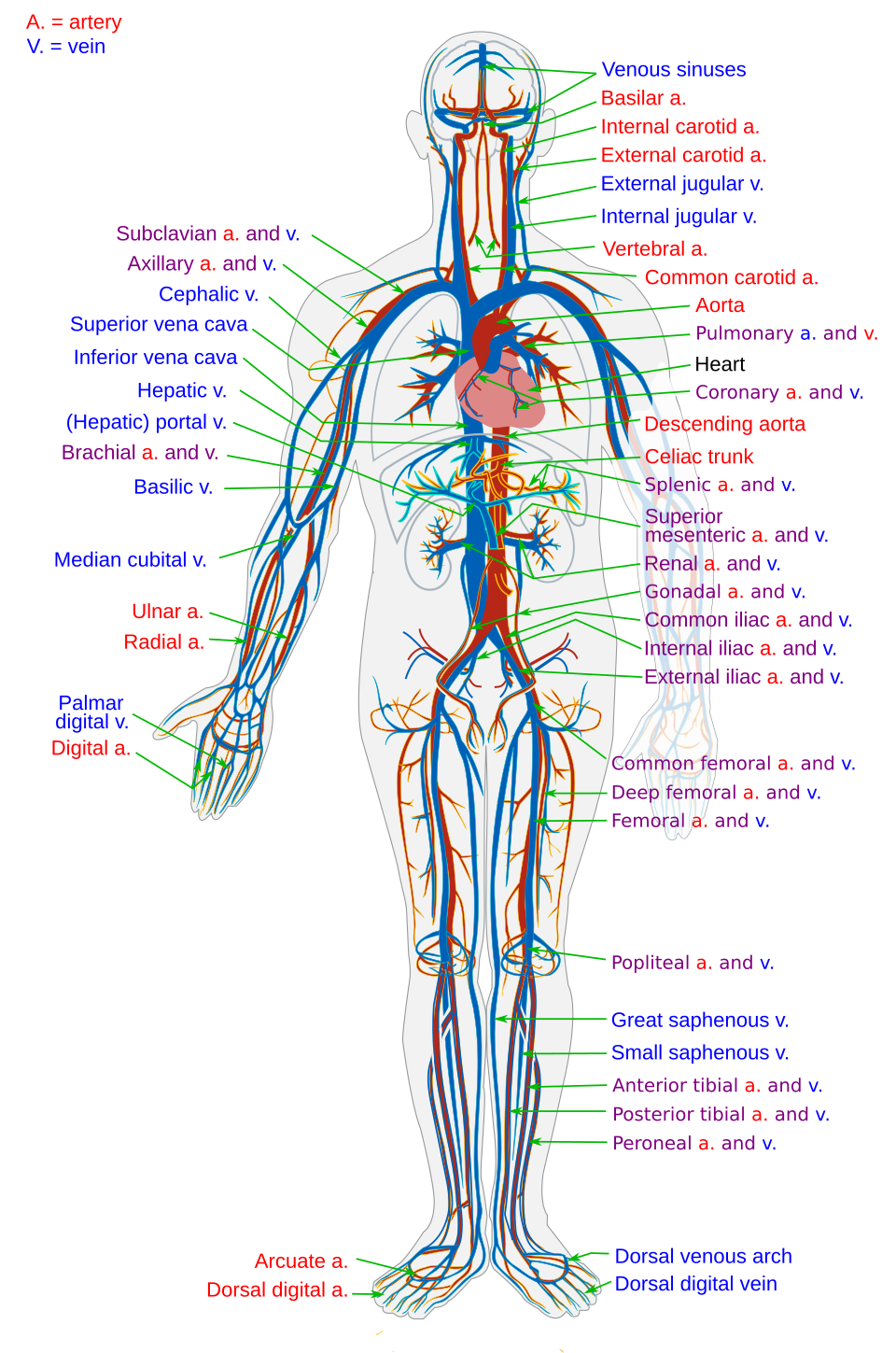
Blood Flow - Closed Double Circulation

Humans have a closed circulatory system - blood stays within blood vessels at all times. Blood flows through two connected circuits (figure-of-8 pattern):

  1. Systemic circuit: oxygenated blood from left ventricle → aorta → arteries → capillaries (exchange in organs) → veins → vena cava → right atrium. Delivers O₂/nutrients, collects CO₂/waste.
  2. Pulmonary circuit: deoxygenated blood from right ventricle → pulmonary artery → lungs (gas exchange) → pulmonary vein → left atrium. Blood circulates through both circuits roughly once per minute.

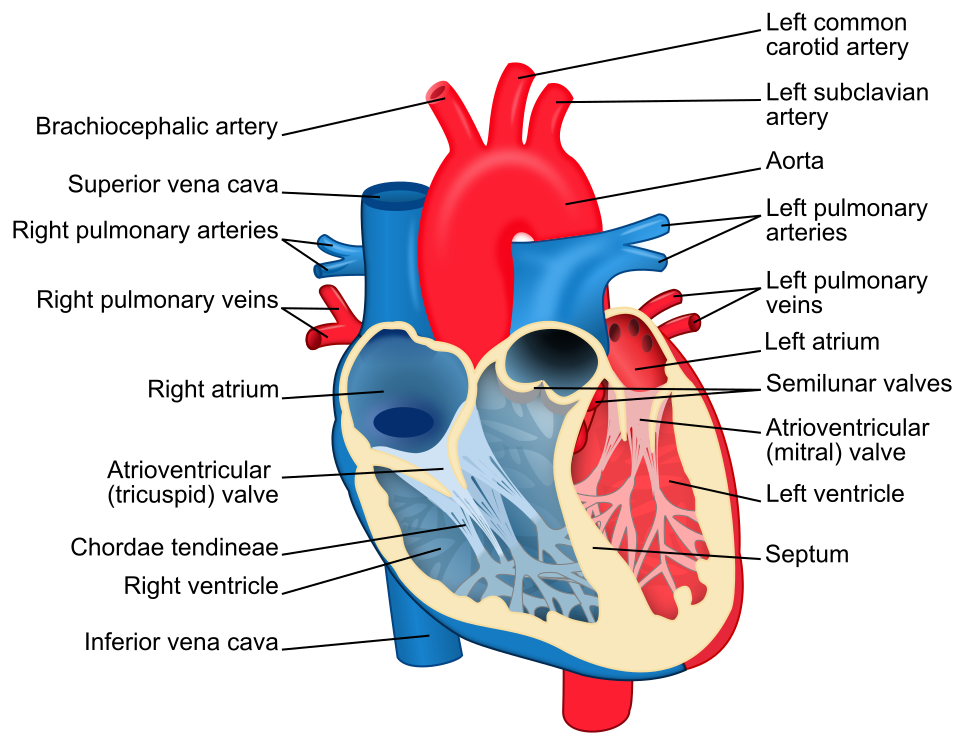
The Heart - Structure

A muscular organ the size of a fist, located behind the breastbone, slightly left of centre. Made of cardiac muscle - does not tire, does not grow, under unconscious control.

4 chambers:

  • Right atrium: receives deoxygenated blood from the body (via vena cava)
  • Right ventricle: pumps blood to the lungs (via pulmonary artery)
  • Left atrium: receives oxygenated blood from lungs (via pulmonary vein)
  • Left ventricle: pumps blood to the body (via aorta) - thickest wall (highest pressure needed)
  • Septum: muscular wall separating left and right sides

Valves prevent backflow: tricuspid (right AV), bicuspid/mitral (left AV), semi-lunar valves (in aorta and pulmonary artery).

Coronary arteries supply the heart muscle itself with oxygenated blood. Blockage of coronary arteries → heart attack.

The Cardiac Cycle

The repeating sequence of changes inside the heart during each heartbeat. Three stages:

  1. Diastole (relaxation): all chambers relax. Blood flows into atria from veins.
  2. Atrial systole: atria contract → push blood through AV valves into ventricles.
  3. Ventricular systole: ventricles contract → push blood out through semi-lunar valves into arteries (aorta and pulmonary artery). AV valves close (prevents backflow).

Pacemaker: the sinoatrial node (SA node/SAN) - a small patch of tissue in the right atrium - generates electrical impulses that initiate each heartbeat. It controls the rhythm of the cardiac cycle. The impulse spreads to the atrioventricular node (AV node) which relays it to the ventricles.

Pulse: the repeating expansion and contraction of artery walls caused by ventricular systole. Felt where arteries are close to the surface (e.g. wrist, neck).

Blood pressure: the force of blood pushing against artery walls. Measured as systolic/diastolic (e.g. 120/80 mmHg). Exercise, stress, diet all affect heart rate and blood pressure.

Blood Vessels

Blood flows: heart → arteryarteriolecapillaryvenulevein → heart.

FeatureArteryVeinCapillary
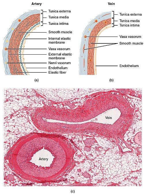
WallThick, muscular, elasticThinner, less muscleOne cell thick
LumenSmallLargeVery narrow (one RBC wide)
ValvesNo (except semi-lunar)Yes (prevent backflow)No
PressureHigh (pulse)Low (no pulse)Very low
DirectionAway from heartTowards heartConnects arterioles to venules
PositionDeep under skinClose to skin surfaceThroughout tissues
Blood typeMostly oxygenatedMostly deoxygenatedExchange zone
MovementPulse pushes bloodMuscle squeeze + valvesDiffusion of materials

Capillary function: walls only one cell thick + vast network = enormous surface area → perfect conditions for diffusion of O₂, nutrients, CO₂ and waste between blood and body cells.

Thermoregulation by Blood Vessels

  • Vasodilation: blood vessels near skin widen → warm blood flows to surface → heat radiates away → skin appears red.
  • Vasoconstriction: blood vessels near skin narrow → less blood near surface → heat conserved → skin appears pale.

The Lymphatic System

When blood passes through capillaries, some plasma leaks out into spaces between cells, forming tissue fluid. The lymphatic system collects this excess fluid:

  • Lymphatic vessels: collect excess tissue fluid (now called lymph) and return it to the circulatory system via the subclavian veins in the upper chest.
  • Lymph nodes: bean-shaped swellings that filter lymph, trapping bacteria and viruses. Contain white blood cells. Swell when fighting infection.
  • Spleen: filters blood, removes old/damaged red blood cells, stores white blood cells.
  • Tonsils: trap pathogens entering via mouth/nose.
  • Thymus: where T-lymphocytes mature.

Interactions with Other Body Systems

  • Breathing system: gas exchange in lungs - CO₂ out, O₂ in via alveoli capillaries.
  • Digestive system: absorbed nutrients enter blood through capillaries in small intestine.
  • Urinary system: waste products (urea) filtered from blood at kidneys.
  • Endocrine system: hormones transported via bloodstream to target organs.
Heart - chambers, valves, vessels

Heart - chambers, valves, vessels

Wikimedia Commons (public domain / CC)

Human circulatory system

Human circulatory system

Wikimedia Commons (public domain / CC)

process

The Cardiac Cycle

Diastole
All chambers relax
Blood fills atria
Atrial Systole
Atria contract
Blood → ventricles
Ventricular Systole
Ventricles contract
Blood → arteries
Cycle repeats
SA node sets rhythm
→ AV node relays
process

Double Circulatory System

Right Ventricle
Lungs
(gas exchange:
CO₂ out, O₂ in)
Left Atrium
→ Left Ventricle
Body organs
(delivers O₂
collects CO₂)
Right Atrium
comparison

Blood Vessel Comparison

Arteries
  • Thick elastic walls
  • Small lumen
  • High pressure (pulse)
  • Away from heart
  • Deep under skin
Veins
  • Thinner walls
  • Large lumen + valves
  • Low pressure
  • Towards heart
  • Near skin surface
Capillaries
  • One cell thick
  • Very narrow lumen
  • Exchange zone
  • Connects arterioles ↔ venules
  • Vast network (diffusion)
diagram

The Lymphatic System

Tissue fluid forms when plasma leaks from capillaries into spaces between cells.

  • Lymphatic vessels collect excess tissue fluid (lymph) → return via subclavian veins
  • Lymph nodes filter lymph, trap pathogens, contain WBCs
  • Spleen filters blood, stores WBCs, removes old RBCs
  • Tonsils trap pathogens at mouth/nose entry
  • Thymus where T-lymphocytes mature

Key Points

  • 1Blood = plasma (55%) + blood cells (45%). Plasma: water, albumin, globulins, fibrinogen, nutrients, waste.
  • 2RBCs: biconcave, no nucleus, haemoglobin binds O₂. WBCs: monocytes (phagocytosis) + lymphocytes (antibodies). Platelets: clotting.
  • 3Closed double circulation: pulmonary circuit (heart → lungs → heart) + systemic circuit (heart → body → heart).
  • 4Heart: 4 chambers, cardiac muscle (never tires), septum, 3 types of valves. Left ventricle thickest (highest pressure).
  • 5Cardiac cycle: diastole (relax) → atrial systole (atria contract) → ventricular systole (ventricles contract). SA node = pacemaker, AV node relays.
  • 6Pulse = artery wall expansion. Blood pressure = force on artery walls (systolic/diastolic).
  • 7Blood flow: artery → arteriole → capillary → venule → vein. Capillaries: one cell thick, vast surface area for diffusion.
  • 8Thermoregulation: vasodilation (cooling) vs vasoconstriction (heat conservation).
  • 9Lymphatic system: lymph vessels collect tissue fluid → lymph nodes filter → returned via subclavian veins. Spleen, tonsils, thymus.
  • 10Blood groups: A, B, AB (universal recipient), O (universal donor). Rhesus factor: Rh+ or Rh−.

Learning Outcomes

  • Investigate the structures of the heart and relate them to their functions, use primary and secondary data to support conclusions
  • Model the interaction between the circulatory and other human body systems in facilitating transport of materials around the body
  • Outline heartbeat and its control by the pacemaker, pulse, blood pressure and the cardiac blood supply
  • Relate the composition of the blood to its functions; appreciate the value of knowledge on blood grouping for human health
  • Distinguish between arteries, veins and capillaries based on their macrostructures and role in the circulatory system of humans

Transport in Plants

In brief:Plants transport water, minerals, and sugars through specialised vascular tissues - xylem and phloem.

Plant Structure for Transport

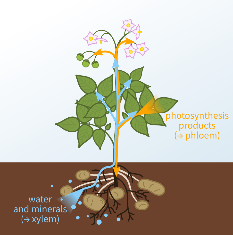
  • Root system: root hairs increase surface area for absorption of water and minerals from soil
  • Root cortex: water moves through cortex cells to reach the vascular tissue
  • Stem: contains vascular bundles (xylem + phloem)
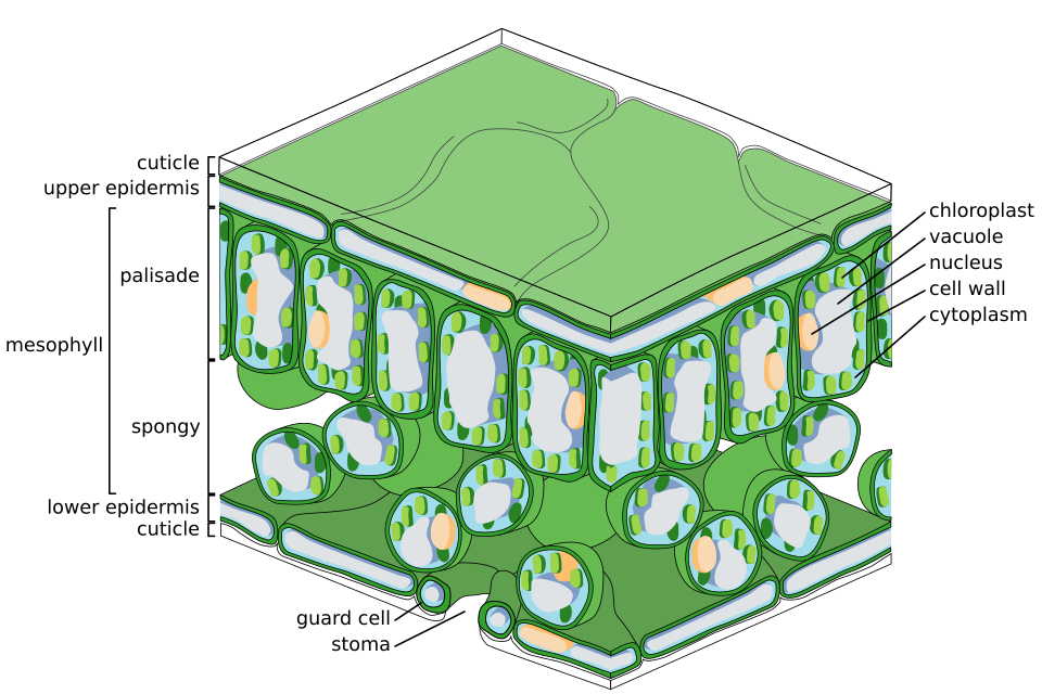
  • Leaves: site of photosynthesis, gas exchange via stomata, and water loss (transpiration)
  • Lenticels: pores in bark that allow gas exchange in woody stems

Xylem - Water and Mineral Transport (Upward)

  • Made of dead cells with thick lignin walls (waterproof, provides structural support)
  • Forms continuous hollow tubes from roots to leaves
  • Water moves upward by:
    • Root pressure (HL): osmosis pushes water into xylem from root hair cells
    • Cohesion-tension (HL): water molecules stick together (cohesion) forming a continuous column. Transpiration from leaves creates a pulling force (tension) that draws water upward
    • Transpiration pull: water evaporating from leaf stomata creates suction

Phloem - Sugar Transport (Translocation)

  • Made of living sieve tube elements with companion cells
  • Transports dissolved sucrose from source (leaves - where photosynthesis occurs) to sink (roots, fruits, growing points)
  • This process is called translocation

Transpiration

Loss of water vapour from leaves through stomata (tiny pores on underside of leaves, controlled by guard cells).

Factors affecting transpiration rate:

  • Air movement (wind): increases rate (removes humid air near leaf)
  • Temperature: increases rate (more evaporation)
  • Light intensity: increases rate (stomata open for photosynthesis)
  • Surface area: more leaves = more transpiration
  • Cuticle: waxy layer reduces water loss
  • CO₂ concentration: high CO₂ → stomata close (less transpiration)
Xylem and phloem tissues

Xylem and phloem tissues

Wikimedia Commons (public domain / CC)

Leaf cross-section

Leaf cross-section

Wikimedia Commons (public domain / CC)

Key Points

  • 1Root hairs absorb water and minerals. Water moves through cortex to xylem.
  • 2Xylem: dead cells, lignin walls. Transports water and minerals upward via cohesion-tension and transpiration pull.
  • 3Phloem: living sieve tubes. Translocates sucrose from source (leaves) to sink (roots, fruits).
  • 4Transpiration: water loss through stomata. Factors: wind, temperature, light, surface area, cuticle, CO₂.
  • 5Root pressure (HL) and cohesion-tension (HL) explain how water rises against gravity in tall plants.

Learning Outcomes

  • Relate the structure of the root, stem and leaf and their associated tissues with their function
  • Explain the transport of water, minerals, carbon dioxide and photosynthetic products in the plant
  • Investigate factors affecting the rate of transpiration in plants, use primary and secondary data to support conclusions

4. Blood - Composition & Function

In brief:Blood is a connective tissue made of cells suspended in plasma. It transports materials, distributes heat and defends against infection.

Blood is roughly 55% plasma and 45% formed elements (cells and platelets). An adult has about 5 litres.

Three main functions

  1. Transport - oxygen, CO₂, nutrients, hormones, urea
  2. Thermoregulation - carries heat produced in muscles/liver around the body
  3. Defence - white blood cells and antibodies fight infection; platelets clot wounds

Plasma

Yellow liquid: ~92% water, ~7% plasma proteins (albumin - maintains pressure/volume; globulins - transport and immunity; fibrinogen - clotting) plus dissolved nutrients, mineral ions and wastes.

Formed elements

CellStructureFunction
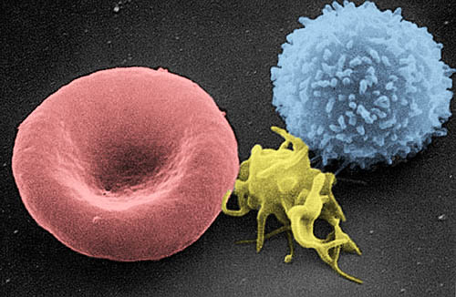
Red blood cellBiconcave disc, no nucleus, packed with haemoglobinTransports O₂ (and some CO₂) around the body
White blood cell - monocyteLarge, single-lobed nucleusEngulfs pathogens by phagocytosis
White blood cell - lymphocyteSmall, round nucleusProduces specific antibodies
PlateletCell fragment, no nucleusTriggers blood clotting at wounds

Blood cell counts can rise or fall in response to infection, altitude or blood loss.

Red and white blood cells

Red blood cells, white blood cells and a platelet in plasma

Blausen Medical / Wikimedia Commons (CC BY)

Key Points

  • 1Blood ≈ 55% plasma + 45% formed elements
  • 2Plasma proteins: albumin (pressure), globulins (immunity/transport), fibrinogen (clotting)
  • 3Red blood cells: biconcave, no nucleus, haemoglobin carries oxygen
  • 4Monocytes engulf pathogens; lymphocytes make antibodies
  • 5Platelets are cell fragments that clot damaged vessels

5. The Heart & Cardiac Cycle

In brief:The heart is a double pump that drives blood through the pulmonary and systemic circuits.

Structure

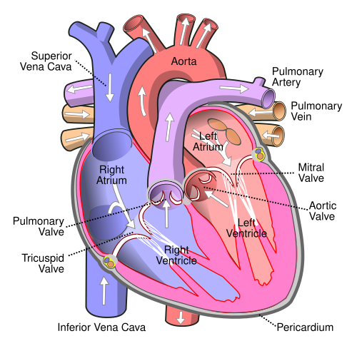
Four chambers: two atria (receive blood) and two ventricles (pump blood out). The septum separates the two sides so oxygenated and deoxygenated blood never mix. Valves stop backflow: bicuspid (left atrium-ventricle), tricuspid (right), and semilunar valves at the exits of the ventricles.

Blood pathway

  1. Body → vena cava → right atrium → right ventricle
  2. Right ventricle → pulmonary artery → lungs (picks up O₂)
  3. Lungs → pulmonary vein → left atrium → left ventricle
  4. Left ventricle → aorta → body

The left ventricle has the thickest muscular wall because it pumps blood to the whole body. Coronary arteries branching from the aorta supply the heart muscle itself with oxygen.

Cardiac cycle (heartbeat)

One full cycle takes about 0.8 s at rest:

  • Diastole - all chambers relax; blood flows into the atria
  • Atrial systole - atria contract, pushing blood into ventricles
  • Ventricular systole - ventricles contract, forcing blood into the arteries

The rhythm is set by the SA node (pacemaker) in the right atrium, passed to the AV node and along Purkinje fibres to the ventricles. An artificial pacemaker can take over if this signal fails.

Pulse & blood pressure

Pulse = the expansion of an artery each time the left ventricle contracts (~72 bpm at rest). Blood pressure is the force blood exerts on artery walls, written systolic/diastolic (~120/80 mmHg). Long-term high blood pressure damages arteries and increases the risk of stroke and heart attack.

Human heart labelled

Labelled diagram of the human heart

Wikimedia Commons (public domain)

Key Points

  • 14 chambers: right atrium/ventricle (deoxygenated), left atrium/ventricle (oxygenated)
  • 2Valves prevent backflow; left ventricle wall is thickest
  • 3Pulmonary circuit → lungs; systemic circuit → body
  • 4Coronary arteries supply the heart muscle itself
  • 5SA node = natural pacemaker; AV node relays to ventricles
  • 6Cardiac cycle: diastole → atrial systole → ventricular systole

6. Blood Vessels & Lymphatic System

In brief:Arteries, veins and capillaries have structures suited to their functions. The lymphatic system drains tissue fluid and supports immunity.

Comparison of vessels

ArteryVeinCapillary
DirectionAway from heartBack to heartLinks arteries & veins
WallThick, muscular, elasticThinner, less muscleOne cell thick
LumenNarrowWideVery narrow (one RBC wide)
PressureHigh & pulsingLowFalls along network
ValvesNone (except at heart)Yes - prevent backflowNone

Arterioles and venules are the small versions that connect arteries and veins to capillary beds. Arterioles carry out vasoconstriction and vasodilation to redirect blood flow (e.g. away from skin when cold).

Capillary exchange

The thin walls and slow flow of blood in capillaries let oxygen, glucose and other small molecules diffuse into tissue fluid, while CO₂ and wastes diffuse back in.

Lymphatic system

Not all tissue fluid returns to capillaries. The excess is picked up by lymphatic vessels and, once inside them, is called lymph. Lymph passes through lymph nodes (bean-shaped filters full of white blood cells that trap bacteria) before draining back into a large vein near the heart. Related organs include the spleen, thymus and tonsils - all involved in immunity.

Artery vs vein structure

Cross-section comparison of an artery, vein and capillary

OpenStax / Wikimedia Commons (CC BY)

Key Points

  • 1Arteries: thick elastic walls, narrow lumen, high pressure, away from heart
  • 2Veins: thinner walls, wide lumen, valves, back to heart
  • 3Capillaries: one cell thick - exchange with tissue fluid
  • 4Vasoconstriction/vasodilation redirect blood flow
  • 5Lymph = tissue fluid inside lymphatic vessels; nodes filter it
  • 6Spleen, thymus and tonsils support immunity

7. The Urinary System

In brief:The kidneys filter blood, remove urea, and control the water and salt balance of the body (osmoregulation).

Excretion is the removal of the waste products of metabolism. Homeostasis is keeping a stable internal environment - the kidneys are central to both.

Urinary system parts

  • Kidneys - bean-shaped organs that filter blood
  • Ureters - carry urine from kidneys to bladder
  • Bladder - stores urine
  • Urethra - releases urine from the body

Blood enters each kidney via the renal artery and leaves - now filtered - via the renal vein. About 5 litres of blood is filtered ~60 times each day.

Regions of the kidney

  1. Capsule - tough outer layer
  2. Cortex - contains the openings of the nephrons; site of filtration
  3. Medulla - contains loops of Henle and collecting ducts
  4. Renal pelvis - funnel that collects urine before it enters the ureter

The nephron (functional unit)

Each kidney holds around a million nephrons. Urine forms in three stages:

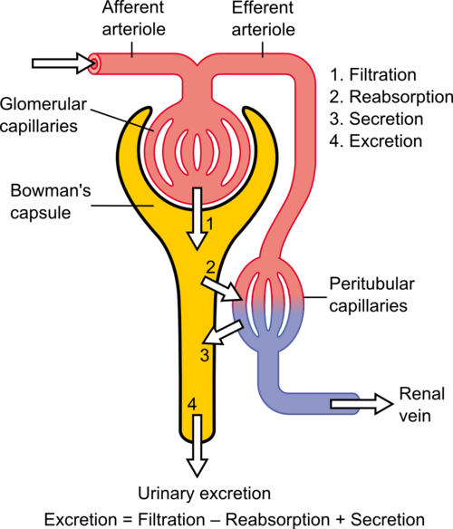
  1. Filtration - blood pushed at high pressure through the glomerulus (a knot of capillaries) into the Bowman's capsule. Water, salts, glucose, urea pass across; blood cells and large proteins stay in the blood. The result is the glomerular filtrate.
  2. Reabsorption - useful materials (glucose, amino acids, most water and salts) are returned to the blood along the proximal tubule and loop of Henle. Glucose uses active transport; water follows by osmosis.
  3. Secretion & concentration - a small amount of extra waste is secreted into the tubule. The distal tubule and collecting duct fine-tune water reabsorption under the hormone ADH (antidiuretic hormone). Dehydration → more ADH → more water reabsorbed → darker, more concentrated urine.

Urea forms in the liver during deamination - the breakdown of excess amino acids. Ammonia (very toxic) is quickly converted to urea before travelling in the blood to the kidney.

Kidney internal structure

Internal structure of the kidney - cortex, medulla, renal pelvis

OpenStax / Wikimedia Commons (CC BY)

Nephron structure and function

Nephron - filtration, reabsorption and secretion

Wikimedia Commons (CC)

Key Points

  • 1Urinary system: kidneys, ureters, bladder, urethra
  • 2Kidney regions: capsule, cortex, medulla, renal pelvis
  • 3Nephron = functional unit; ~1 million per kidney
  • 4Filtration in glomerulus; reabsorption in proximal tubule + loop of Henle
  • 5ADH controls how much water is reabsorbed in the distal tubule
  • 6Urea is made in the liver from excess amino acids (deamination)